Advanced Quantitative Fluorescence Microscopy to Probe the Molecular Dynamics of Viral Entry, Science Lab
Por um escritor misterioso
Descrição
Viral entry into the host cell requires the coordination of many cellular and viral proteins in a precise order. Modern microscopy techniques are now allowing researchers to investigate these interactions with higher spatiotemporal resolution than ever before. Here we present two examples from the field of HIV research that make use of an innovative quantitative imaging approach as well as cutting edge fluorescence lifetime-based confocal microscopy methods to gain novel insights into how HIV fuses to cell membranes and enters the cell.
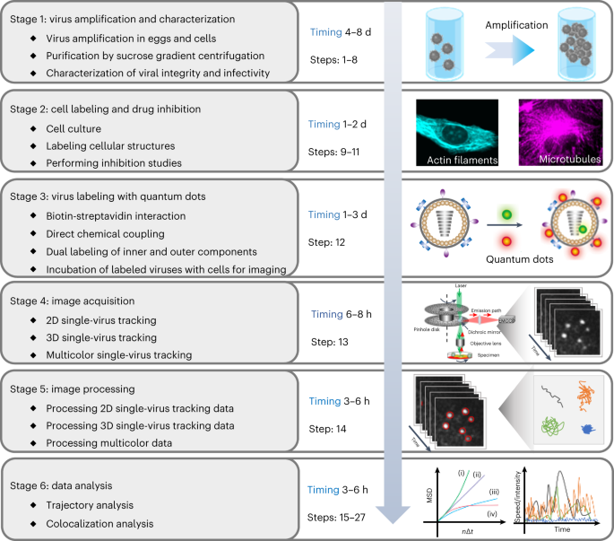
Single-virus tracking with quantum dots in live cells
Frontiers Detecting and measuring of GPCR signaling – comparison of human induced pluripotent stem cells and immortal cell lines
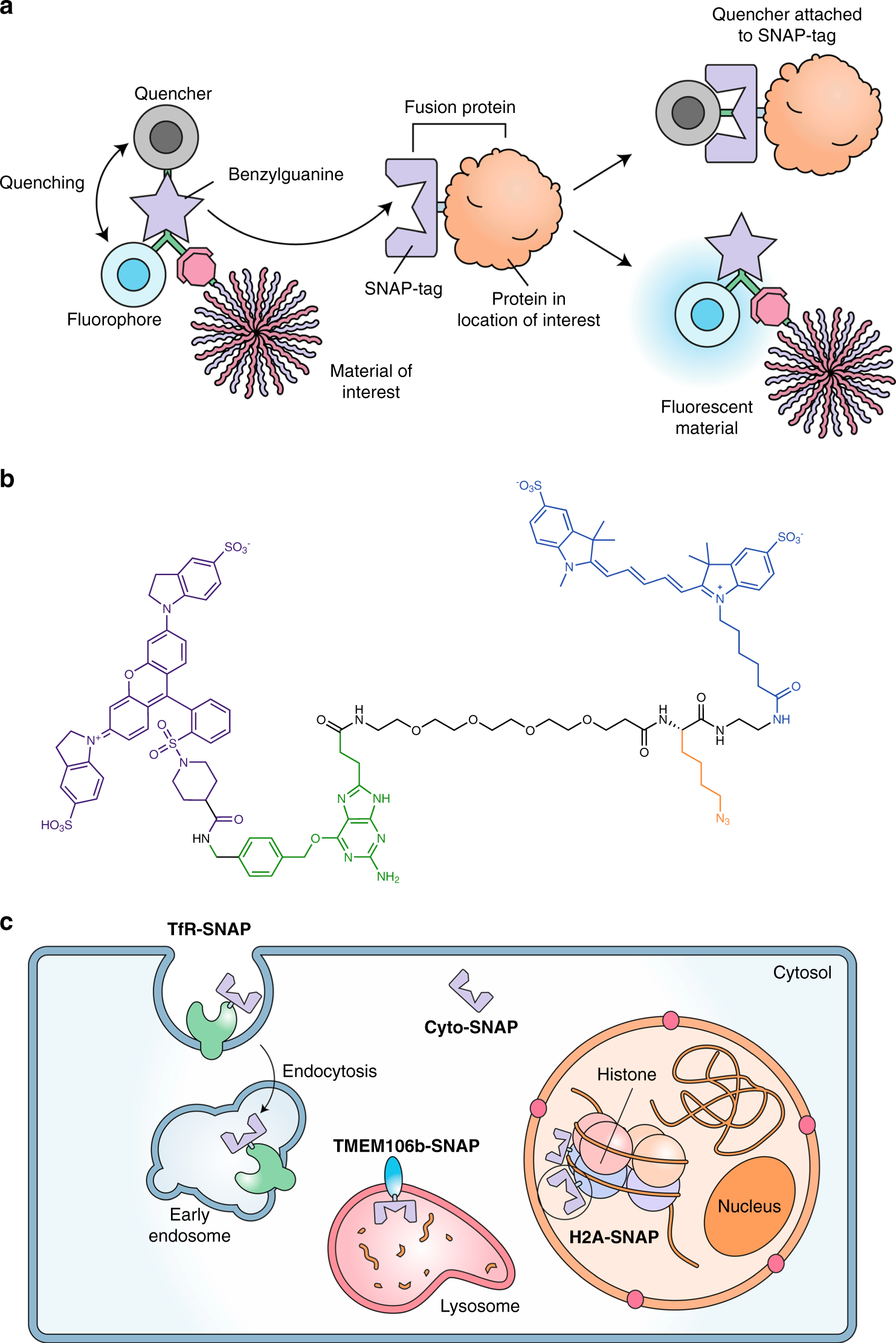
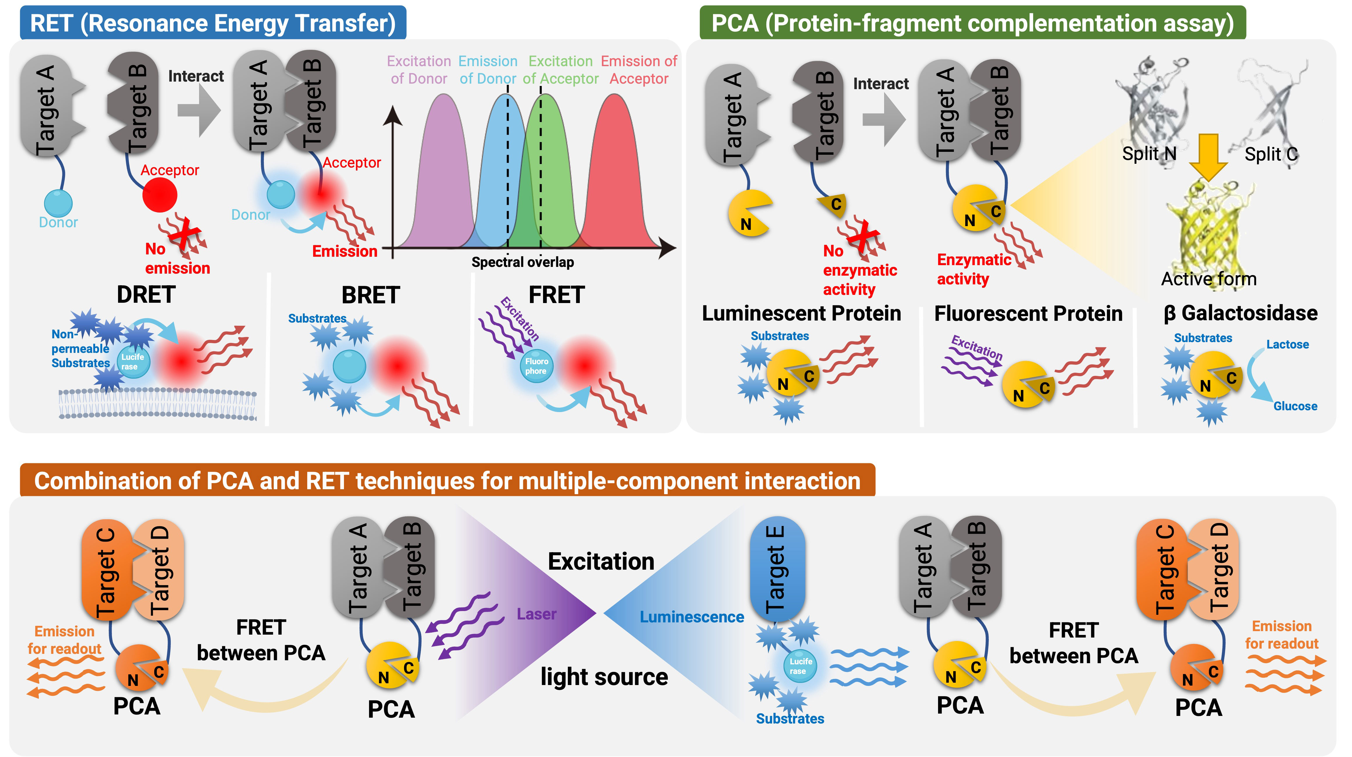
A molecular sensor to quantify the localization of proteins, DNA and nanoparticles in cells
Microscopy in Virology, Science Lab

A chimeric virus-based probe unambiguously detects live circulating tumor cells with high specificity and sensitivity - ScienceDirect
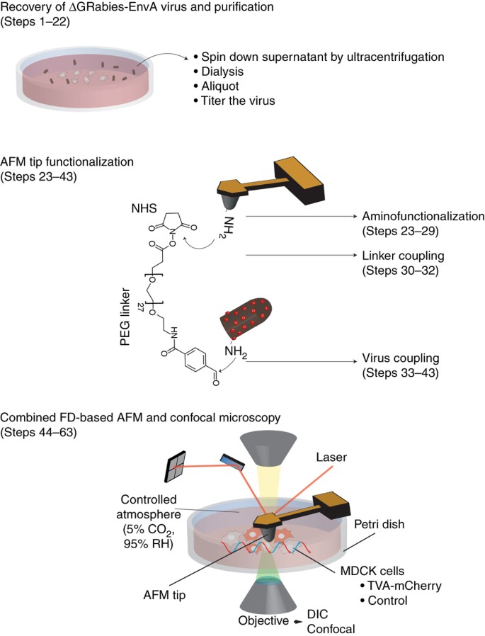
Combining confocal and atomic force microscopy to quantify single-virus binding to mammalian cell surfaces

Advancements in detection of SARS-CoV-2 infection for confronting COVID-19 pandemics - Laboratory Investigation

CRISPR‐Cas Biochemistry and CRISPR‐Based Molecular Diagnostics - Weng - 2023 - Angewandte Chemie International Edition - Wiley Online Library

Influenza A virus exploits transferrin receptor recycling to enter host cells
Viruses, Free Full-Text

Single-Virus Tracking: From Imaging Methodologies to Virological Applications

HIV-1 binding and fusion events observed using confocal fluorescence
de
por adulto (o preço varia de acordo com o tamanho do grupo)